If you've ever experienced a sharp, stabbing pain in your heel with your first steps in the morning, you're likely familiar with plantar fasciitis, a condition that affects approximately 2 million Americans annually and accounts for up to 15% of all foot-related complaints requiring professional care.
At Podoks, we understand the profound impact this condition can have on your daily life, athletic pursuits, and overall well-being.
This comprehensive guide explores the symptoms, causes, and evidence-based treatments for plantar fasciitis, including how biomechanically designed socks can play a supportive role in management and prevention.
By understanding this common but complex condition, you'll be better equipped to address it effectively and return to pain-free movement.
What is Plantar Fasciitis?
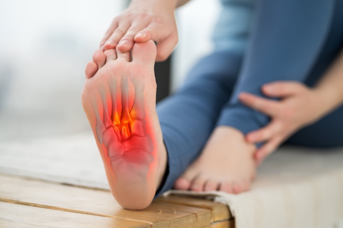
Plantar fasciitis is an inflammatory condition affecting the plantar fascia; a thick, fibrous band of connective tissue that runs along the bottom of the foot, connecting the heel bone (calcaneus) to the toes and creating the arch of the foot. This critical structure functions as a shock-absorbing bowstring that supports the arch and assists with proper foot mechanics during walking, running, and standing.
When excessive stress causes small tears in the plantar fascia, the body responds with inflammation and irritation, leading to the characteristic pain and stiffness associated with plantar fasciitis. While often described as an inflammatory condition, recent research suggests that many chronic cases actually involve degenerative changes to the fascia rather than acute inflammation, leading some medical professionals to use the term "plantar fasciosis" for long-standing cases (Lemont et al., 2003).

Recognizing the Symptoms
Plantar fasciitis presents with several distinctive symptoms that help differentiate it from other foot conditions:
Classic Morning Pain
The hallmark symptom is sharp, stabbing pain localized to the inside front of the heel, particularly with the first few steps after waking. This characteristic "first-step pain" occurs because the plantar fascia tightens overnight. When you suddenly place weight on the contracted tissue in the morning, it creates tension that triggers pain at the fascia's attachment to the heel bone.
This pain typically decreases after the first few minutes of walking as the fascia gradually stretches, though it may return after prolonged periods of sitting or standing.
Pain Patterns Throughout the Day
Unlike some orthopedic conditions that cause consistent pain, plantar fasciitis pain often follows a characteristic pattern: improvement after initial morning stiffness, followed by increasing discomfort with extended weight-bearing activities.
Many patients report that pain intensifies toward the end of the day, particularly after long periods of standing or after completing exercise, rather than during the activity itself.
Tenderness to Touch
Direct pressure on the medial tubercle of the calcaneus (the inside front portion of the heel bone) typically elicits significant tenderness in plantar fasciitis patients.
This specific location, where the plantar fascia attaches to the heel bone—is the most common site of pain and inflammation.
Limited Ankle Motion
Restricted upward movement of the foot at the ankle (dorsiflexion) frequently accompanies plantar fasciitis due to tightness in the calf muscles and Achilles tendon. This limitation can be assessed by attempting to pull the toes toward the shin while keeping the knee straight.

Common Causes and Risk Factors
Understanding the underlying causes and risk factors for plantar fasciitis can help both prevent initial development and avoid recurrence after treatment. Research has identified several significant contributors:
Biomechanical Factors
Foot structure and mechanics play crucial roles in plantar fasciitis development:
Excessive Pronation: When feet roll inward excessively during the gait cycle, the arch flattens more than optimal, placing additional tension on the plantar fascia. Research has shown that individuals with excessive pronation experience significantly higher rates of plantar fasciitis compared to those with neutral mechanics.
High Arches (Pes Cavus): Very high, rigid arches also increase plantar fasciitis risk. These structures provide less natural shock absorption, transferring more impact forces to the plantar fascia. Clinical studies have demonstrated that individuals with high arches had a 2.4 times greater risk of developing plantar fasciitis.
Leg Length Discrepancy: Even minor differences in leg length can create asymmetrical loading patterns that place excess stress on the plantar fascia of the more heavily loaded foot.
Activity-Related Factors
How we use our feet significantly influences plantar fasciitis development:
Sudden Increases in Activity: Rapidly increasing mileage, intensity, or duration of weight-bearing activities without proper adaptation time overwhelms the plantar fascia's ability to recover between sessions. This pattern explains the common occurrence among runners who significantly increase their training volume too quickly.
Surface Changes: Transitioning from softer to harder running or walking surfaces without adequate adaptation time increases impact forces transmitted to the plantar fascia.
Occupational Demands: Professions requiring prolonged standing or walking, particularly on hard surfaces, create cumulative stress on the plantar fascia. Studies show that healthcare workers, teachers, factory workers, and retail employees have significantly higher plantar fasciitis rates compared to sedentary occupations.
Footwear Considerations
The shoes we choose dramatically affect foot function and plantar fascia stress:
Inadequate Support: Shoes lacking proper arch support force the plantar fascia to bear excessive load without assistance, particularly during prolonged standing or walking.
Worn Footwear: Athletic shoes lose their cushioning capacity after extended use, even when they appear visually intact. This degradation increases impact forces reaching the plantar fascia.
Inappropriate Shoe Types: Regularly wearing completely flat shoes or shoes with excessive heel heights creates unnatural loading patterns that stress the plantar fascia.
Physical and Demographic Factors
Certain physical characteristics influence plantar fasciitis susceptibility:
Age: Incidence peaks between ages 40-60, reflecting age-related changes in fascia elasticity and fat pad thinning under the heel.
Weight: Research demonstrates that individuals with body mass index (BMI) greater than 30 have significantly higher rates of plantar fasciitis compared to those with lower BMI measurements.
Calf Tightness: Tight gastrocnemius and soleus muscles create increased tension on the Achilles tendon, which indirectly transfers stress to the connected plantar fascia (19).

Diagnosis: Confirming Plantar Fasciitis
While the symptoms often strongly suggest plantar fasciitis, proper diagnosis requires professional evaluation to rule out other possible conditions with similar presentations.
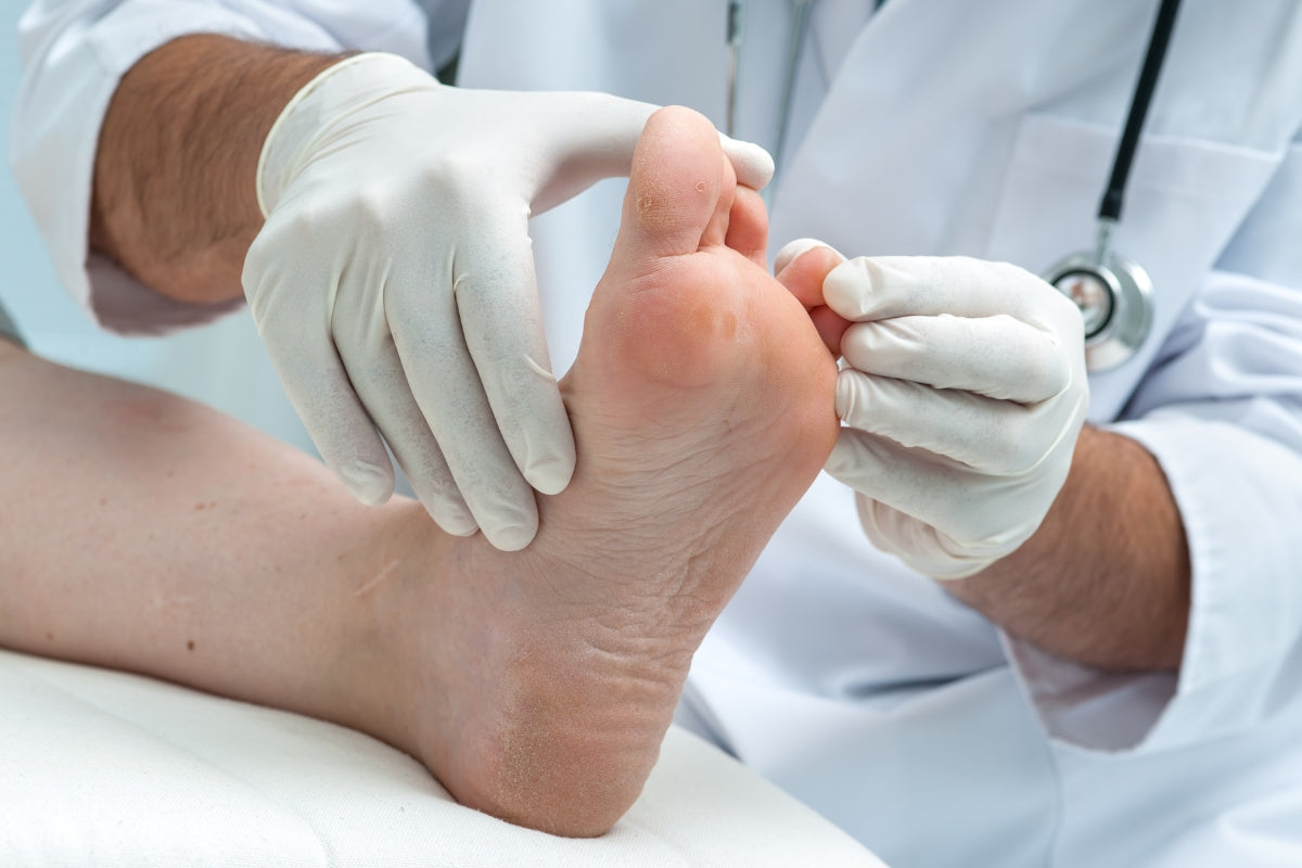
Clinical Examination
A thorough physical examination by a healthcare provider typically includes:
- Palpation of the plantar fascia to identify specific pain locations.
- Assessment of foot structure and arch height.
- Evaluation of ankle range of motion, particularly dorsiflexion.
- Observation of gait patterns and foot mechanics.
The presence of tenderness at the medial calcaneal tubercle, positive windlass test (pain with toe extension), and characteristic symptom patterns provide strong diagnostic evidence.
Imaging Studies
While plantar fasciitis remains primarily a clinical diagnosis, imaging may help rule out other conditions or assess severity:
Ultrasonography: Particularly useful for plantar fasciitis, ultrasound can visualize fascia thickening (>4mm is considered abnormal) and detect areas of degeneration or tears.
X-rays: While not showing the plantar fascia itself, x-rays can identify heel spurs (present in approximately 50% of plantar fasciitis cases) and rule out stress fractures or other bony abnormalities.
MRI: Typically reserved for cases considering surgical intervention or when alternative diagnoses are strongly suspected, MRI provides detailed visualization of soft tissue structures.
Evidence-Based Treatment Approaches
Effective plantar fasciitis management typically involves a multi-modal approach addressing both symptoms and underlying causes:
First-Line Treatments
Research consistently supports several initial interventions:
Rest and Activity Modification: Temporarily reducing activities that aggravate symptoms, particularly high-impact exercises, allows damaged tissue to begin healing. Cross-training with lower-impact activities like swimming or cycling can maintain fitness during this period.
Ice Therapy: Applying ice to the painful area for 15-20 minutes several times daily, particularly after activity, helps manage pain and inflammation.
Stretching Protocols: Consistent stretching of the plantar fascia and calf muscles represents one of the most effective interventions. Research by DiGiovanni et al. found that structured stretching programs produced significant improvement in a majority of patients within 8-12 weeks.
The most effective stretch involves pulling the toes backward toward the shin while keeping the knee straight, creating tension along the entire posterior chain. Holding this position for 30 seconds and repeating 3-5 times, several sessions daily, provides cumulative benefit.
Supportive Footwear: Shoes with proper arch support, cushioned heels, and adequate shock absorption significantly reduce symptoms during recovery and help prevent recurrence. Research has shown that appropriate supportive footwear combined with stretching exercises provided better outcomes than stretching alone.
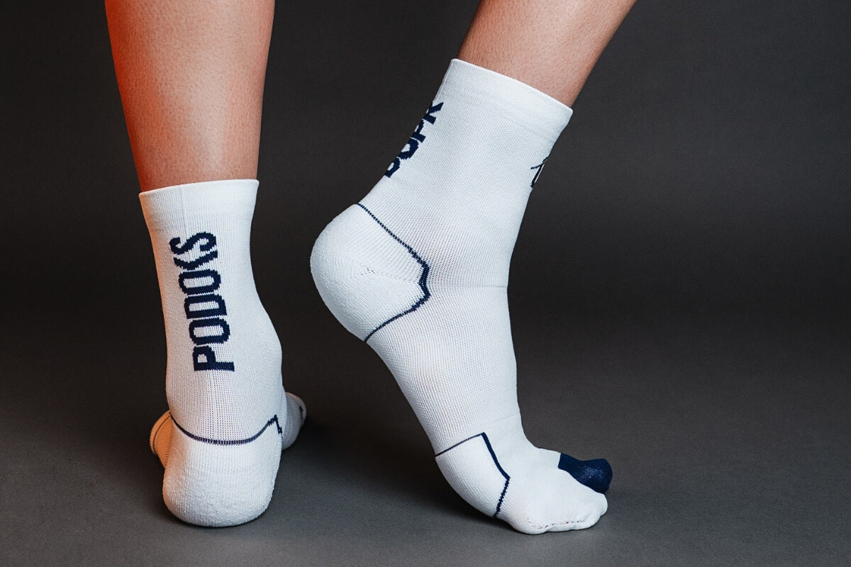
Properly designed biomechanical socks may represent a valuable component of comprehensive plantar fasciitis care.
Specialized Support Options
Beyond basic approaches, several specialized support options have demonstrated effectiveness:
Night Splints: These devices maintain a gentle stretch on the plantar fascia and calf muscles during sleep, preventing the overnight tightening that causes morning pain. Controlled trials have found that night splints combined with stretching produced significantly better outcomes than stretching alone, particularly for reducing morning pain.
Orthotic Devices: Custom or high-quality over-the-counter orthotics provide arch support that reduces tension on the plantar fascia. Systematic reviews have found that orthotics effectively reduced pain in a majority of patients within 6-12 weeks of consistent use.
The Role of Biomechanical Socks
Recent research has investigated the potential benefits of specially designed biomechanical socks in plantar fasciitis management. Studies by Martínez-Nova et al. demonstrated that biomechanical socks can reduce plantar pressure under the first metatarsal head by 25% and under the first toe by 23%, potentially alleviating stress on the plantar fascia.
Additionally, research by Gómez-Carrión et al. found that biomechanical socks with reinforced arch support reduced the force required for proper toe dorsiflexion during the windlass mechanism, suggesting improved foot biomechanics.
While these studies show promising results, biomechanical socks should be considered as a supportive adjunct to, rather than a replacement for, established treatments. They may be particularly beneficial when used alongside other conservative measures such as stretching, appropriate footwear, and activity modification.
Medical Interventions
When conservative measures prove insufficient, medical professionals may recommend:
Physical Therapy: Structured programs combining manual therapy, specialized exercises, and education about body mechanics can address both symptoms and underlying causes. Reviews show that formalized physical therapy programs produced significant improvements in 70-80% of plantar fasciitis cases.
Corticosteroid Injections: These anti-inflammatory injections can provide significant temporary relief. However, the effect typically diminishes after 4-12 weeks, and repeated injections may increase the risk of plantar fascia rupture.
Extracorporeal Shockwave Therapy (ESWT): This non-invasive procedure uses sound waves to stimulate healing in chronic cases. Meta-analyses have found ESWT effective for approximately 50% of patients who hadn't responded to other conservative treatments.
Platelet-Rich Plasma (PRP) Injections: This emerging therapy uses the patient's own growth factors to potentially accelerate healing. Controlled trials have demonstrated that PRP injections provided better long-term outcomes than corticosteroid injections for chronic plantar fasciitis.
Surgical Options
Reserved for severe cases unresponsive to at least 6-12 months of conservative treatment, surgical interventions include:
Plantar Fascia Release: Partially detaching the plantar fascia from the heel bone to relieve tension. Long-term studies have found success rates between 70-90% for carefully selected surgical candidates.
Gastrocnemius Recession: Lengthening the calf muscles to reduce pull on the plantar fascia. Research has demonstrated that this procedure, either alone or in combination with plantar fascia release, provided good to excellent results in 75-85% of cases.
Surgery is considered only after exhausting other options due to potential complications and variable outcomes.

Prevention Strategies: Avoiding Recurrence
Once recovered from plantar fasciitis, preventing recurrence becomes the priority. Research indicates that approximately 30% of individuals who recover will experience another episode without proper preventive measures.
These strategies help maintain long-term foot health:
Gradual Activity Progression
Adhering to the 10% rule when increasing activity levels allows tissues to adapt appropriately to increasing demands. This principle applies to running mileage, walking distance, or standing duration.
Consistent Stretching Routine
Maintaining calf and plantar fascia flexibility through daily stretching prevents the tightness that often precedes symptom recurrence. Long-term follow-up studies found that continuing a regular stretching program after symptom resolution reduced recurrence risk by approximately 60%.
Footwear Vigilance
Replacing athletic shoes every 300-500 miles and avoiding unsupportive casual footwear helps maintain proper foot support. Prospective studies found that regular footwear rotation and timely replacement significantly reduced injury rates among runners.
Weight Management
Since excess weight dramatically increases forces through the plantar fascia, maintaining healthy body weight represents one of the most effective prevention strategies for those with previous plantar fasciitis episodes.
Ongoing Support
According to research, continuing to wear supportive devices like biomechanical socks during high-risk activities may provide protection against recurring damage. While more research is needed, early evidence suggests that continued use of properly designed supportive footwear may help prevent recurrence.

Conclusion: A Path Forward from Plantar Fasciitis
While plantar fasciitis can significantly impact quality of life, the evidence clearly demonstrates that most cases respond well to appropriate treatment. By combining traditional approaches like stretching and activity modification with emerging supportive technologies like biomechanical socks, the vast majority of individuals can achieve lasting relief.
Understanding both the underlying causes and evidence-based treatments empowers you to take control of your recovery process. Whether you're currently managing plantar fasciitis or seeking to prevent its development, implementing the strategies outlined in this guide provides a clear path toward healthy, pain-free feet.
At Podoks, we remain committed to applying cutting-edge research to create products that genuinely enhance foot health. While properly designed biomechanical socks may represent a valuable component of comprehensive plantar fasciitis care, they work best as part of a complete treatment approach that includes appropriate medical care, activity modification, and other proven interventions.
------
Scientific References:
Buchanan BK, Sina RE, Kushner D. Plantar Fasciitis. StatPearls [Internet]. Treasure Island (FL): StatPearls Publishing; 2024. PMID: 28613664
Martin RL, Davenport TE, Reischl SF, et al. Heel pain—plantar fasciitis: revision 2014. J Orthop Sports Phys Ther. 2014;44(11):A1-A33. doi:10.2519/jospt.2014.0303
Thomas JL, Christensen JC, Kravitz SR, et al. The diagnosis and treatment of heel pain: a clinical practice guideline–revision 2010. J Foot Ankle Surg. 2010;49(3):S1-S19. doi:10.1053/j.jfas.2010.01.001
Lemont H, Ammirati KM, Usen N. Plantar fasciitis: a degenerative process (fasciosis) without inflammation. J Am Podiatr Med Assoc. 2003;93(3):234-237. doi:10.7547/87507315-93-3-234
Wearing SC, Smeathers JE, Urry SR, Hennig EM, Hills AP. The pathomechanics of plantar fasciitis. Sports Med. 2006;36(7):585-611. doi:10.2165/00007256-200636070-00004
Cole C, Seto C, Gazewood J. Plantar fasciitis: evidence-based review of diagnosis and therapy. Am Fam Physician. 2005;72(11):2237-2242. PMID:16342847
Bartold SJ. The plantar fascia as a source of pain-biomechanics, presentation and treatment. J Bodyw Mov Ther. 2004;8(3):214-226. doi:10.1016/j.jbmt.2004.01.006
Patel A, DiGiovanni B. Association between plantar fasciitis and isolated contracture of the gastrocnemius. Foot Ankle Int. 2011;32(1):5-8. doi:10.3113/FAI.2011.0005
Irving DB, Cook JL, Young MA, Menz HB. Obesity and pronated foot type may increase the risk of chronic plantar heel pain: a matched case-control study. BMC Musculoskelet Disord. 2007;8:41. doi:10.1186/1471-2474-8-41
Riddle DL, Pulisic M, Pidcoe P, Johnson RE. Risk factors for plantar fasciitis: a matched case-control study. J Bone Joint Surg Am. 2003;85(5):872-877. doi:10.2106/00004623-200305000-00015
Brady RJ, Dean JB, Skinner TM, Gross MT. Limb length inequality: clinical implications for assessment and intervention. J Orthop Sports Phys Ther. 2003;33(5):221-234. doi:10.2519/jospt.2003.33.5.221
Di Caprio F, Buda R, Mosca M, et al. Foot and lower limb diseases in runners: assessment of risk factors. J Sports Sci Med. 2010;9(4):587-596. PMID:24149785
Ferris DP, Liang K, Farley CT. Runners adjust leg stiffness for their first step on a new running surface. J Biomech. 1999;32(8):787-794. doi:10.1016/S0021-9290(99)00078-0
Werner RA, Gell N, Hartigan A, et al. Risk factors for plantar fasciitis among assembly plant workers. PM R. 2010;2(2):110-116. doi:10.1016/j.pmrj.2010.01.001
Rajan P, Anderson J. Choosing proper footwear. StatPearls [Internet]. Treasure Island (FL): StatPearls Publishing; 2022. PMID:32310560
Cook SD, Kester MA, Brunet ME. Shock absorption characteristics of running shoes. Am J Sports Med. 1985;13(4):248-253. doi:10.1177/036354658501300406
Schwartz EN, Su J. Plantar fasciitis: a concise review. Perm J. 2014;18(1):e105-e107. doi:10.7812/TPP/13-113
Taunton JE, Ryan MB, Clement DB, et al. A retrospective case-control analysis of 2002 running injuries. Br J Sports Med. 2002;36(2):95-101. doi:10.1136/bjsm.36.2.95
McMillan AM, Landorf KB, Barrett JT, et al. Diagnostic imaging for chronic plantar heel pain: a systematic review and meta-analysis. J Foot Ankle Res. 2013;6:20. doi:10.1186/1757-1146-6-20
Jeswani T, Morlese J, McNally EG. Getting to the heel of the problem: plantar fascia lesions. Clin Radiol. 2008;63(11):1254-1263. doi:10.1016/j.crad.2008.01.016
Buchbinder R. Plantar fasciitis. N Engl J Med. 2004;350(21):2159-2166. doi:10.1056/NEJMcp032745
DiGiovanni BF, Nawoczenski DA, Lintal ME, et al. Tissue-specific plantar fascia-stretching exercise enhances outcomes in patients with chronic heel pain. A prospective, randomized study. J Bone Joint Surg Am. 2003;85(7):1270-1277. doi:10.2106/00004623-200307000-00013
DiGiovanni BF, Nawoczenski DA, Malay DP, et al. Plantar fascia-specific stretching exercise improves outcomes in patients with chronic plantar fasciitis. A prospective clinical trial with two-year follow-up. J Bone Joint Surg Am. 2006;88(8):1775-1781. doi:10.2106/JBJS.E.01281
Ryan M, Fraser S, McDonald K, Taunton J. Examining the degree of pain reduction using a multielement exercise model with a conventional training shoe versus an ultraflexible training shoe for treating plantar fasciitis. Phys Sportsmed. 2014;37(4):68-74. doi:10.3810/psm.2009.12.1746
Beyzadeoglu T, Gokce A, Bekler H. The effectiveness of dorsiflexion night splint added to conservative treatment for plantar fasciitis. Acta Orthop Traumatol Turc. 2007;41(3):220-224. PMID:17876118
Landorf KB, Keenan AM, Herbert RD. Effectiveness of foot orthoses to treat plantar fasciitis: a randomized trial. Arch Intern Med. 2006;166(12):1305-1310. doi:10.1001/archinte.166.12.1305
Martínez-Nova A, González-Alonso A, Fernández-Miranda Gastón M, Morán Cortés JF. Reducción de presiones plantares dinámicas en el antepié plantar medial con calcetines biomecánicos. Rev Esp Pod. 2022;33(2):87-92. doi:10.20986/revesppod.2022.1650/2022
Gómez-Carrión Á, Reguera-Medina JM, Coheña-Jiménez M, et al. Biomechanical Effect on Jack's Test on Barefoot Position, Regular Socks, and Biomechanics Socks. Life (Basel). 2024;14(2):248. doi:10.3390/life14020248
David JA, Sankarapandian V, Christopher PR, et al. Injected corticosteroids for treating plantar heel pain in adults. Cochrane Database Syst Rev. 2017;6:CD009348. doi:10.1002/14651858.CD009348.pub2
Yin MC, Ye J, Yao M, et al. Is extracorporeal shock wave therapy clinical efficacy for relief of chronic, recalcitrant plantar fasciitis? A systematic review and meta-analysis of randomized placebo or active-treatment controlled trials. Arch Phys Med Rehabil. 2020;101(8):1451-1460. doi:10.1016/j.apmr.2020.03.013
Mahindra P, Yamin M, Selhi HS, et al. Chronic plantar fasciitis: effect of platelet-rich plasma, corticosteroid, and placebo. Orthopedics. 2016;39(2):e285-e289. doi:10.3928/01477447-20160222-01
Davies MS, Weiss GA, Saxby TS. Plantar fasciitis: how successful is surgical intervention? Foot Ankle Int. 1999;20(12):803-807. doi:10.1177/107110079902001207
Abbassian A, Kohls-Gatzoulis J, Solan MC. Proximal medial gastrocnemius release in the treatment of recalcitrant plantar fasciitis. Foot Ankle Int. 2012;33(1):14-19. doi:10.3113/FAI.2012.0014
Gill LH, Kiebzak GM. Outcome of nonsurgical treatment for plantar fasciitis. Foot Ankle Int. 1996;17(9):527-532. doi:10.1177/107110079601700903
Nielsen RO, Buist I, Sørensen H, et al. Training errors and running related injuries: a systematic review. Int J Sports Phys Ther. 2012;7(1):58-75. PMID:22319684
Taunton JE, Ryan MB, Clement DB, et al. A prospective study of running injuries: the Vancouver Sun Run "In Training" clinics. Br J Sports Med. 2003;37(3):239-244. doi:10.1136/bjsm.37.3.239
Leave a comment
This site is protected by hCaptcha and the hCaptcha Privacy Policy and Terms of Service apply.